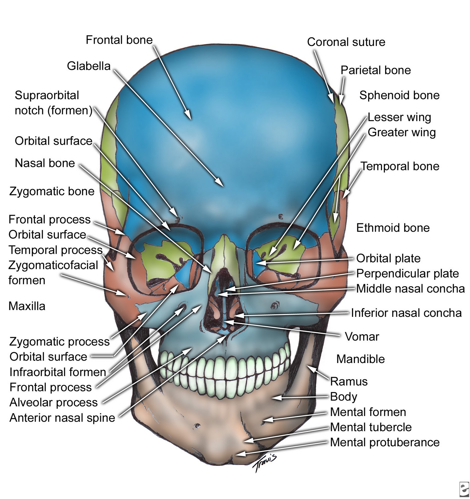
Decoding the Dome: A Deep Dive into Skull Anatomy Imagery
Ever feel like you're carrying the weight of the world on your shoulders? Well, technically, you're carrying it slightly higher – on your skull! This intricate bony structure, more than just a helmet for your brain, is a complex marvel of evolutionary design. And thanks to the magic of imaging, we can delve into its depths without, you know, any actual digging.
Visual representations of the skull, from ancient anatomical drawings to high-tech 3D models, have always captivated and informed. They’re not just morbid curiosities; they’re windows into our very being. Think about it: skull imagery unlocks a secret language written in bone, telling stories of evolution, health, and even artistic expression.
The journey of skull depictions begins centuries ago, with sketches etched onto papyrus and cave walls. These early attempts, while rudimentary, demonstrate a primal fascination with the skeletal framework that defines us. Fast forward through history, and the study of skull anatomy illustrations becomes integral to medical understanding, driving advancements in surgery and diagnostics. Illustrations in medical texts evolved from basic outlines to detailed cross-sections, reflecting an ever-deepening grasp of cranial complexity.
Today, skull imagery takes many forms. We've got everything from classic anatomical charts used in classrooms to interactive digital models that let you rotate and zoom in on every foramen and suture. These visual tools are vital for medical professionals, researchers, and anyone with a thirst for knowledge about the human body. The ability to visualize the intricate network of bones, sinuses, and foramina is critical for diagnosing fractures, tumors, and other cranial conditions.
But the significance of skull visuals extends beyond the purely scientific. Skull motifs pop up everywhere, from edgy fashion statements to artistic expressions exploring themes of mortality and identity. This pervasive presence in popular culture speaks to our complex relationship with this potent symbol.
Medical professionals use different types of skull imagery, like X-rays, CT scans, and MRI scans, depending on the diagnostic needs. An X-ray provides a 2D view, helpful for spotting fractures. A CT scan offers more detailed cross-sectional images for identifying bone abnormalities. An MRI scan visualizes soft tissues within the skull, crucial for examining the brain.
Benefits of skull images include accurate diagnosis of head injuries, planning complex surgeries, and educating patients about their conditions. For example, a surgeon can use a 3D skull model generated from a CT scan to plan the best approach for removing a tumor, minimizing risk and maximizing precision.
Advantages and Disadvantages of Skull Imagery
| Advantages | Disadvantages |
|---|---|
| Accurate diagnosis | Radiation exposure (in some cases) |
| Surgical planning | Cost of advanced imaging |
| Educational tool | Potential for misinterpretation |
Five Best Practices: 1. Use high-quality images from reputable sources. 2. Always consider the context and purpose of the image. 3. Consult with medical professionals for interpretation when necessary. 4. Properly label and cite images in academic or professional settings. 5. Be mindful of cultural sensitivity when using skull imagery in non-medical contexts.
Real Examples: 1. CT scans used to diagnose skull fractures. 2. 3D printed skull models used for surgical planning. 3. Anatomical charts used in medical education. 4. Skull imagery in art and design. 5. Forensic anthropology using skull analysis for identification.
FAQ: 1. What does a skull fracture look like on an X-ray? 2. How are 3D skull models created? 3. What are the different bones of the skull? 4. How can I learn more about skull anatomy? 5. What is the role of skull imagery in forensic science? 6. What are the ethical considerations of using skull imagery? 7. What are some common misconceptions about skull anatomy? 8. Where can I find reliable resources for skull anatomy images?
Tips and Tricks: When studying skull anatomy images, focus on landmarks like sutures and foramina. Use interactive models to explore the 3D structure. Compare different types of images (X-ray, CT, MRI) to understand their respective strengths. Always consult reputable sources for accurate information.
In conclusion, from medical marvels to cultural mainstays, representations of the human skull continue to intrigue and inform. Whether you're a medical professional, a budding artist, or simply curious about the skeletal structure that safeguards your thoughts and dreams, delving into skull anatomy imagery offers a unique and valuable perspective. These visual tools are not just pictures; they are portals into the complex and fascinating landscape of the human body. So, next time you see a skull, take a moment to appreciate the wealth of information it holds – a testament to our shared human story etched in bone. Understanding our own anatomy empowers us to take better care of ourselves, appreciate the intricacies of our existence, and perhaps, carry the weight of the world a little more lightly.
Ryan reynolds wrexham film more than just a hollywood story
Setting sail smartly your guide to the best boat loan rates near you
Fresh bengali fish delivered find your nearest market


